Dental radiographs, commonly referred to as X-ray films, or informally, X-rays, are pictures of the teeth, bones, and surrounding soft tissues to screen for and help identify problems with the teeth, mouth, and jaw.
X-ray pictures can show cavities, cancerous or benign masses, hidden dental structures (such as wisdom teeth), and bone loss that cannot be seen during a visual examination. Dental X-rays may also be done as follow-up after dental treatments.
A radiographic image is formed by a controlled burst of X-ray radiation which penetrates oral structures at different levels, depending on varying anatomical densities, before striking the film or sensor. Teeth appear lighter because less radiation penetrates them to reach the film. Dental caries, infections and other changes in the bone density, and the periodontal ligament, appear darker because X-rays readily penetrate these less dense structures. Dental restorations (fillings, crowns) may appear lighter or darker, depending on the density of the material.
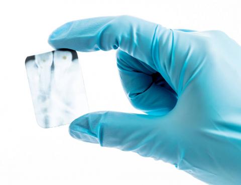
Sansone Dental Practice has invested in digital radiography so that our patients receive only minimal exposure to radiation compared to what they received using x-ray film.
This preoperative photo of tooth #3, (A), reveals no clinically apparent decay other than a small spot within the central fossa. In fact, decay could not be detected with an explorer. Radiographic evaluation, (B), however, revealed an extensive region of demineralization within the dentin (arrows) of the mesial half of the tooth. When a bur was used to remove the occlusal enamel overlying the decay, (C), a large hollow was found within the crown and it was discovered that a hole in the side of the tooth, was contiguous with this hollow. After all of the decay had been removed, (D), the pulp chamber had been exposed and most of the mesial half of the crown was either missing or poorly supported.
It is possible for both tooth decay and periodontal disease not to be visible during a clinical exam, and radiographic evaluation of the dental and periodontal tissues is a critical segment of the comprehensive oral examination. The photographic montage above depicts a situation in which extensive decay could not be detected prior to radiographic evaluation.